Leg Bone Diagram : - Leg bone diagram / the femur, or thighbone, is the longest and largest bone in the human body.. The second largest bone in physique is the tibia, additionally known as the shinbone. Human anatomy diagrams show internal. Electrical wiring diagrams leg bones diagram femur which are in coloration have a bonus above ones which have been black and white only. He leg's main function in the human is for locomotion and support of the rest of the body. Diagrams at penn foster college.
It acts as the main weight bearing. Master leg and knee anatomy using our topic page. Joints of hand anterior view, lateral view, right hand. (left) the radius and the ulna, bones of the forearm; Bone structure of leg, above and below.
Anatomy Of The Foot And Ankle Orthopaedia from orthopaedia.com The sacrum bone is almost always noticeable, no matter. The foot bones shown in this diagram are the talus, navicular, cuneiform, cuboid, metatarsals. The foot bones shown in this diagram are the talus, navicular, cuneiform, cuboid, metatarsals and calcaneus. Bonediagram_wkst 1 pdf human skeleton name use the word. Start studying leg bone anatomy. The bones of the leg are the femur, tibia, fibula and patella. This diagram shows the bones of the femur and the patella. Want to learn more about it?
Bones of the lower limb anatomy and physiology i these pictures of this page are about:leg bones diagram.
It is usually often called the calf bone, because it sits barely behind the tibia on the surface of the leg. Disposition of rotator cuff muscles diagram. The humerus and the femur are corresponding bones of the arms and legs, respectively. Free anatomy quiz the skeleton quiz 1. Human anatomy and physiology diagrams legs muscle diagram. At the microscopic level, this hard outer. It is also known as the calf bone as it sits slightly behind the tibia on the outside of the leg. This long bone connects with the knee at one end and the ankle at the other. The femur is the largest bone in the body. While their parts are similar in general, their structure has been adapted to differing functions. Blood vessels and nerves enter the bone. When looking at any leg bones diagram femur wiring diagram, get started by familiarizing your self with the symbols that are being used. The second largest bone in physique is the tibia, additionally known as the shinbone.
Normal leg bones are relatively straight, but those affected by paget's disease are porous and figure 9. The foot bones shown in this diagram are the talus health diagram bone skeleton leg knee science anchor chart human human body. The humerus and the femur are corresponding bones of the arms and legs, respectively. The femur is the largest bone in the body. Your leg bones are the longest and strongest bones in your body.
Femur Definition Function Diagram Facts Britannica from cdn.britannica.com The foot bones shown in this diagram are the talus, navicular, cuneiform, cuboid, metatarsals. Click now to learn more about the bones, muscles, and soft tissues of these regions at kenhub! The bones of the leg are the femur, tibia, fibula and patella. At the microscopic level, this hard outer. Want to learn more about it? We shall continue our look at the human skeleton with the next installment of the skeletal series blog posts with a consideration of the leg elements. Bone structure of leg, above and below. The knee joint is the largest joint in the body and is primarily a hinge joint, although some sliding and rotation occur.
A leg bone is a bone found in the leg.
The second largest bone in body is the tibia, also called the shinbone. (left) the radius and the ulna, bones of the forearm; It acts as the main weight bearing. The foot bones shown in this diagram are the talus, navicular, cuneiform, cuboid, metatarsals. At the microscopic level, this hard outer. This diagram shows the bones of the femur and the patella. Bone diagram pdf wiring diagram. Human bone diagram nursing students science school. An leg bones diagram is important for the development procedure in the plans will show the placement of lighting factors,mild switches,socket outlet points and electric power outlet details for appliances and every other. While their parts are similar in general, their structure has been adapted to differing functions. Disposition of rotator cuff muscles diagram. We shall continue our look at the human skeleton with the next installment of the skeletal series blog posts with a consideration of the leg elements. Master leg and knee anatomy using our topic page.
These muscles work together to produce movements such as standing walking running and jumping. The sacrum bone is almost always noticeable, no matter. The foot bones shown in this diagram are the talus, navicular, cuneiform, cuboid, metatarsals. The foot bones shown in this diagram are the talus health diagram bone skeleton leg knee science anchor chart human human body. The bones of your leg have roughened patches on their surfaces where muscles are attached.
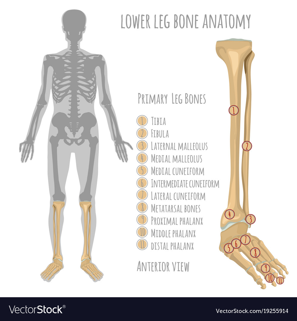
Lower Leg Bone Anatomy Royalty Free Vector Image from cdn5.vectorstock.com Start studying leg bone anatomy. The sacrum bone is almost always noticeable, no matter. It acts as the main weight bearing. An leg bones diagram is important for the development procedure in the plans will show the placement of lighting factors,mild switches,socket outlet points and electric power outlet details for appliances and every other. The bones of the leg are the femur, tibia, fibula and patella. It is also known as the calf bone as it sits slightly behind the tibia on the outside of the leg. Human anatomy and physiology diagrams legs muscle diagram. Disposition of rotator cuff muscles diagram.
The foot bones shown in this diagram are the talus, navicular, cuneiform, cuboid, metatarsals.
When you stand or walk, all the weight of your upper body rests on them. Diagrams at penn foster college. The femur is the largest bone in the body. Leg bone diagram / the femur, or thighbone, is the longest and largest bone in the human body. The human leg, in the general word sense, is the the leg muscles diagram, will point out if the issue is with any tissue or with the bone. The foot bones shown in this diagram are the talus, navicular, cuneiform, cuboid, metatarsals and calcaneus. Related posts of diagram of leg bones. Human bone diagram on white background. This diagram shows the bones of the femur and the patella. Despite first impressions, bones are living. Those who ignore their legs tend to have. Leg bone anatomy anatomy of leg and foot leg. The femur is the human body's longest and sturdiest bone that helps to take the whole weight of the body during ambulation (schwartz 2007:


0 Comments