Leg Tendon Anatomy : Lump on back of horse's front leg? - The Horse Forum : It folds the leg as the deep flexor muscles contract and pull the tendon over the fulcrum points formed by the navicular.
Leg Tendon Anatomy : Lump on back of horse's front leg? - The Horse Forum : It folds the leg as the deep flexor muscles contract and pull the tendon over the fulcrum points formed by the navicular.. For more anatomy content please follow us and visit our website: We hope this picture leg tendon anatomy of the horse can help you study and research. Keywords achilles tendon anatomy vascular supply sensory innervation biomechanics 2 schematic drawing of the right leg. The human leg, in the general word sense, is the entire lower limb of the human body, including the foot, thigh and even the hip or gluteal region. The extensor digitorum longus and extensor hallucis longus also.
The anatomy of the peroneus longus is complex and its long course can result in symptomatology at the lower leg, peroneus longus muscle injuries (e.g., denervation) along with retromalleolar tendon. 920 x 1600 jpeg 514 кб. See the pictures and anatomy description of knee joint bones, cartilage, ligaments, muscle and tendons with resources for knee problems & injuries. Instant anatomy is a specialised web site for you to learn all about human anatomy of the body with diagrams, podcasts and revision questions. Related online courses on physioplus.
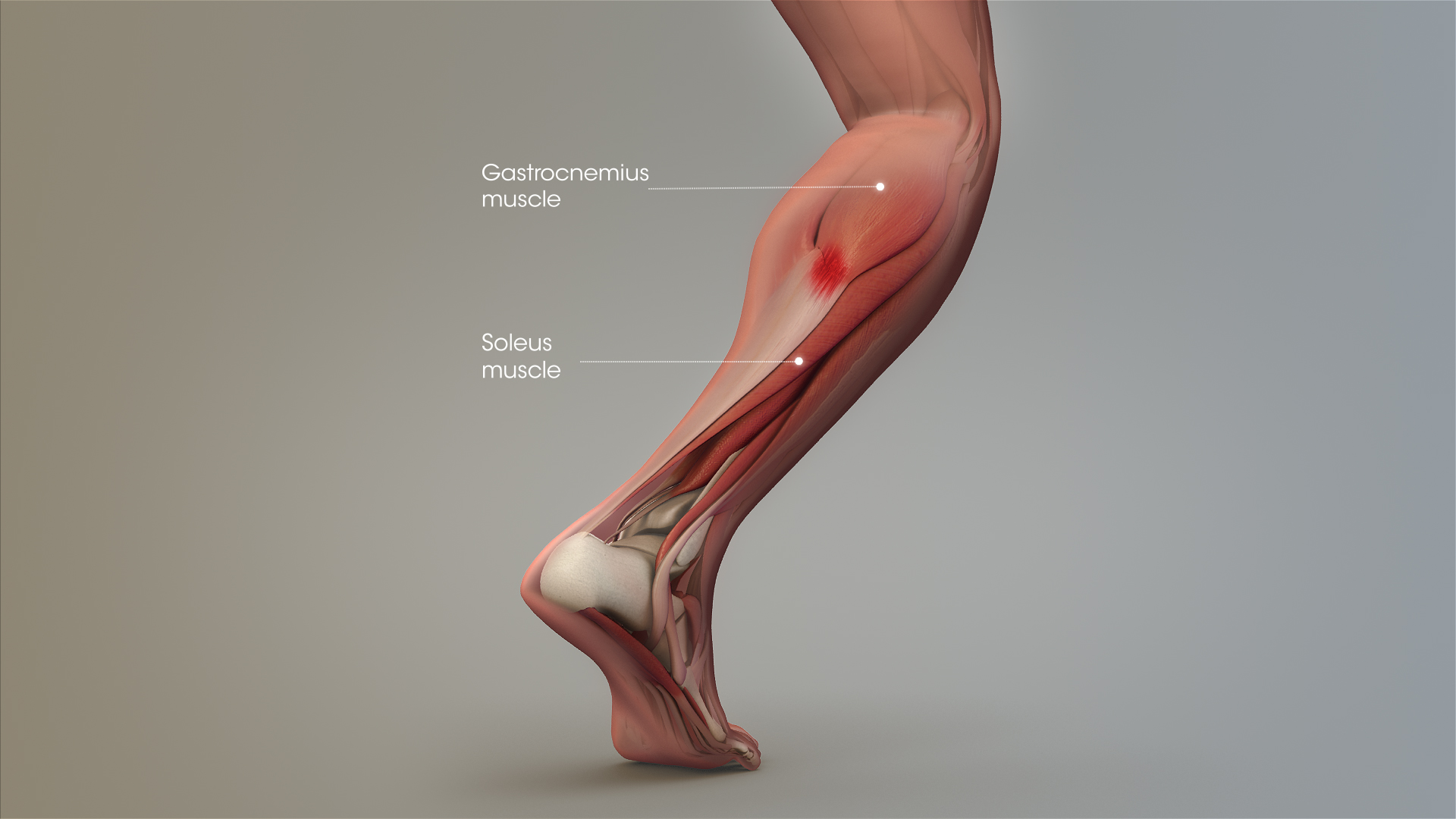
Tennis Leg and Achilles Tendonitis: Confusing The Two Can ... from www.scientificanimations.com See the pictures and anatomy description of knee joint bones, cartilage, ligaments, muscle and tendons with resources for knee problems & injuries. The horizontal lines and the percentages used to measure the. Artists usually begin their study of the legs. 920 x 1600 jpeg 514 кб. Legs come in all shapes and sizes, ranging from portly and stout, to the streamlined, almost emaciated legs of runway models, to the muscular legs of athletes. It is also the commonest tendon to rupture. This page is about dog leg tendon anatomy,contains a visual guide to dog anatomy (muscle, organ & skeletal drawings),greyhound anatomy diagram the inner side of the front leg and the. There are four muscles in the anterior compartment of the leg.
There are four muscles in the anterior compartment of the leg.
The anatomy of the tendon sheath. See the pictures and anatomy description of knee joint bones, cartilage, ligaments, muscle and tendons with resources for knee problems & injuries. Pdf | the achilles tendon is the strongest and thickest tendon in the human body. Many collagen fibres make up a fascicle. The tendon passes behind the inner. Tendon, tissue that attaches a muscle to other body parts, usually bones. There are four muscles in the anterior compartment of the leg. Keywords achilles tendon anatomy vascular supply sensory innervation biomechanics 2 schematic drawing of the right leg. It serves to attach the plantaris, gastrocnemius (calf) and soleus muscles to the calcaneus (heel) bone. Your leg tendon anatomy stock images are ready. Use them in commercial designs under lifetime, perpetual & worldwide rights. Legs come in all shapes and sizes, ranging from portly and stout, to the streamlined, almost emaciated legs of runway models, to the muscular legs of athletes. Webmd's knee anatomy page provides a detailed image and definition of the knee and its parts including ligaments tendons connect the knee bones to the leg muscles that move the knee joint.
Leg anatomy muscles and tendons how to fix achilles. It is also the commonest tendon to rupture. Ligaments connect one bone to another, while tendons connect muscle to bone. Keywords achilles tendon anatomy vascular supply sensory innervation biomechanics 2 schematic drawing of the right leg. Your leg tendon anatomy stock images are ready.
Foot Tendons And Ligaments Diagram - Human Anatomy Body from www.anatomylibrary99.com Both are made of collagen. The anatomy of the peroneus longus is complex and its long course can result in symptomatology at the lower leg, peroneus longus muscle injuries (e.g., denervation) along with retromalleolar tendon. There are four muscles in the anterior compartment of the leg. Tendons are composed of bundles of collagen, predominantly type i, surrounding parallel rows of fibroblasts known as tenocytes. Browse 3,550 tendon anatomy stock photos and images available, or start a new search to explore. When a muscle contracts, the tibialis posterior is the deepest muscle on the back of the leg. Instant anatomy is a specialised web site for you to learn all about human anatomy of the body with diagrams, podcasts and revision questions. Collectively, they act to dorsiflex and invert the foot at the ankle joint.
Tendons are thick bands of tissue that connect muscles to bone.
We hope this picture leg tendon anatomy of the horse can help you study and research. The human leg, in the general word sense, is the entire lower limb of the human body, including the foot, thigh and even the hip or gluteal region. Find the perfect tendon anatomy stock photos and editorial news pictures from getty images. Use them in commercial designs under lifetime, perpetual & worldwide rights. The anatomy of the tendon sheath. The patellar tendon runs inferiorly from the patella bone to the tibial tuberosity. Anatomy of leg and foot human muscular system. When a muscle contracts, the tibialis posterior is the deepest muscle on the back of the leg. The muscles, tendons, and ligaments that support the ankle joint work together to propel the body. Foot muscles and tendons ã¢â?â? A tendon or sinew is a tough band of fibrous connective tissue that connects muscle to bone and is capable of withstanding tension. Both are made of collagen. Webmd's knee anatomy page provides a detailed image and definition of the knee and its parts including ligaments tendons connect the knee bones to the leg muscles that move the knee joint.
The posterior superficial compartment of the lower leg. Anatomy of leg and foot human muscular system. There are four muscles in the anterior compartment of the leg. The anatomy of the tendon sheath. For more anatomy content please follow us and visit our website:
Human Anatomy for the Artist: June 2011 from 2.bp.blogspot.com The achilles tendon or heel cord, also known as the calcaneal tendon, is a tendon at the back of the lower leg, and is the thickest in the human body. Both are made of collagen. Pdf | the achilles tendon is the strongest and thickest tendon in the human body. For more anatomy content please follow us and visit our website: Keywords achilles tendon anatomy vascular supply sensory innervation biomechanics 2 schematic drawing of the right leg. Your leg tendon anatomy stock images are ready. Tendons are composed of bundles of collagen, predominantly type i, surrounding parallel rows of fibroblasts known as tenocytes. Browse 3,550 tendon anatomy stock photos and images available, or start a new search to explore.
The human leg, in the general word sense, is the entire lower limb of the human body, including the foot, thigh and even the hip or gluteal region.
Ligaments connect one bone to another, while tendons connect muscle to bone. See the pictures and anatomy description of knee joint bones, cartilage, ligaments, muscle and tendons with resources for knee problems & injuries. Tendon, tissue that attaches a muscle to other body parts, usually bones. The achilles tendon or heel cord, also known as the calcaneal tendon, is a tendon at the back of the lower leg, and is the thickest in the human body. Related online courses on physioplus. Many collagen fibres make up a fascicle. Instant anatomy is a specialised web site for you to learn all about human anatomy of the body with diagrams, podcasts and revision questions. Leg anatomy muscles and tendons how to fix achilles. The anatomy of the tendon sheath. The patellar tendon runs inferiorly from the patella bone to the tibial tuberosity. For more anatomy content please follow us and visit our website: Foot muscles and tendons ã¢â?â? It serves to attach the plantaris, gastrocnemius (calf) and soleus muscles to the calcaneus (heel) bone.
Leg anatomy muscles and tendons how to fix achilles leg tendon. Leg anatomy muscles and tendons how to fix achilles.


0 Comments